
Retinal Detachment Treatment in Lucknow
Retinal Detachment Treatment in Lucknow is focused on diagnosing retinal separation at the earliest stage and providing appropriate treatment to protect vision. Retinal detachment is a serious eye condition in which the retina separates from the underlying tissue that supports its normal function. Because the retina is responsible for converting light into signals for the brain, a detached retina can affect vision significantly.
At Mansarovar Eye Hospital, patients can receive specialized retinal evaluation, diagnostic testing, and treatment planning for retinal conditions. If you experience sudden flashes of light, a sudden increase in floaters, blurred vision, or a curtain-like shadow in your field of vision, an urgent eye examination is important.
What Is Retinal Detachment?
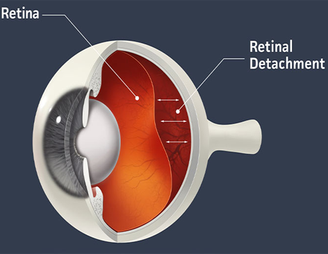
Retinal detachment occurs when the retina pulls away from its normal position at the back of the eye. The retina contains light-sensitive cells that help create the visual information needed for clear sight. When the retina becomes detached, its normal function can be disrupted.
Retinal detachment can develop suddenly or progress over time. In many cases, early symptoms may provide an important warning before significant vision loss occurs. Therefore, recognizing symptoms and seeking prompt medical attention can be important for protecting vision.
The condition can affect one or both eyes, although having retinal detachment in one eye may increase the risk of developing a retinal problem in the other eye.
Symptoms of Retinal Detachment
Knowing the warning signs of retinal detachment can help patients seek timely eye care. Common symptoms may include:
- Sudden appearance of many new floaters
- Flashes of light in the affected eye
- Blurred or distorted vision
- A dark shadow in the peripheral vision
- A curtain-like effect moving across the field of vision
- Reduced side vision
- Sudden changes in visual clarity
Retinal detachment does not always cause pain. This means that the absence of eye pain does not necessarily mean the condition is harmless.
If you suddenly notice flashes, numerous new floaters, or a curtain or shadow over your vision, seek an urgent retinal examination.
What Causes Retinal Detachment?
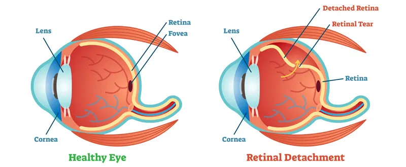
Several factors can contribute to retinal detachment. The most common mechanism involves a retinal tear or break that allows fluid to pass underneath the retina.
Common causes and risk factors include:
Age-Related Changes
As people age, the vitreous gel inside the eye naturally changes. In some cases, these changes can pull on the retina and cause a retinal tear, which may lead to retinal detachment.
Retinal Tears or Breaks
A retinal tear can allow fluid to move underneath the retina. If untreated, this may progress to retinal detachment.
Eye Injury
A significant eye injury can damage the retina or surrounding structures and increase the risk of retinal detachment.
Previous Eye Surgery
Certain eye surgeries may be associated with an increased risk of retinal complications. Regular follow-up examinations can help identify problems early.
Severe Nearsightedness
People with high myopia may have structural changes in the eye that increase the risk of retinal tears and detachment.
Previous Retinal Detachment
A history of retinal detachment in one eye may increase the risk of retinal detachment in the other eye.
Family History
Some individuals may have a higher risk because of inherited eye conditions or a family history of retinal problems.
Types of Retinal Detachment
Retinal detachment can be classified into different types depending on how the retina separates from the underlying tissue.
Rhegmatogenous Retinal Detachment
This is the most common type. It usually occurs when a retinal tear or break allows fluid to pass underneath the retina.
Tractional Retinal Detachment
Tractional retinal detachment occurs when scar tissue or abnormal tissue pulls on the retina. It may occur in association with certain retinal diseases, including advanced diabetic eye disease.
Exudative Retinal Detachment
Exudative retinal detachment occurs when fluid accumulates beneath the retina without a retinal tear or break being the primary cause. It can be associated with inflammation, vascular problems, tumors, or other eye conditions.
Determining the type and cause of retinal detachment is important because treatment depends on the underlying problem.
Diagnosis of Retinal Detachment
A detailed retinal examination is essential when retinal detachment is suspected. An ophthalmologist may examine the retina using specialized instruments and diagnostic techniques.
Depending on the condition, evaluation may include:
- Dilated retinal examination
- Visual acuity testing
- Peripheral retinal examination
- Optical coherence tomography when appropriate
- Ocular ultrasound when the retina cannot be clearly visualized
- Other retinal imaging or diagnostic tests when required
The exact tests depend on the symptoms, clinical findings, and condition of the eye.
Early diagnosis can help the ophthalmologist determine whether immediate treatment is required and which treatment approach may be appropriate.
Retinal Detachment Treatment in Lucknow
The appropriate Retinal Detachment Treatment in Lucknow depends on factors such as the location and size of the detachment, whether a retinal tear is present, how much of the retina is affected, and the overall condition of the eye.
Treatment may include laser procedures, cryotherapy, pneumatic retinopexy, scleral buckle surgery, vitrectomy, or a combination of procedures depending on the individual case.
Laser Treatment for Retinal Tears
If a retinal tear or hole is identified before a significant detachment develops, laser treatment may sometimes be used to create a protective barrier around the affected area.
Laser photocoagulation is performed by an ophthalmologist and is intended to reduce the risk of fluid passing through the retinal break and causing further separation.
Cryotherapy
Cryotherapy uses controlled cold treatment to create a scar around a retinal tear. This scar can help seal the affected area and may be used in selected cases.
Pneumatic Retinopexy
Pneumatic retinopexy involves placing a gas bubble inside the eye. The bubble can help reposition the detached retina against the wall of the eye. Laser treatment or cryotherapy may also be used to seal the retinal break.
This technique is appropriate only for certain retinal detachments.
Scleral Buckle Surgery
A scleral buckle is a surgical procedure in which a flexible band is placed around the outside of the eye. It helps reduce the pulling force on the retina and supports the area containing the retinal tear.
The buckle may remain in place permanently and is generally not visible from outside the eye.
Vitrectomy
Vitrectomy is a retinal surgery in which the vitreous gel is removed from inside the eye. This allows the retinal surgeon to access the retina and treat retinal tears or other abnormalities.
A gas bubble or silicone oil may be used to support the retina while it heals. The specific approach depends on the retinal condition.
Why Timely Retinal Detachment Treatment Matters
Retinal detachment can threaten vision, particularly when the central part of the retina is affected. Delaying evaluation may allow the detachment to progress.
Timely assessment can help determine:
- Whether a retinal tear is present
- Whether the retina is detached
- Whether the macula is involved
- How extensive the detachment is
- Which treatment is appropriate
- Whether urgent retinal surgery is required
For this reason, sudden retinal symptoms should not be ignored.
Who Is at Higher Risk of Retinal Detachment?
Certain people may have a greater risk of retinal detachment. Risk factors may include:
- High myopia
- Previous cataract or other eye surgery
- Previous retinal detachment
- Retinal tears or lattice degeneration
- Significant eye trauma
- Certain inherited eye conditions
- Family history of retinal detachment
- Certain retinal diseases
Having a risk factor does not mean retinal detachment will definitely occur. However, individuals at higher risk may benefit from appropriate retinal examinations.
Retinal Detachment and Floaters
Floaters are small spots, lines, or cobweb-like shapes that appear to move across your vision. They can occur due to normal changes in the vitreous as people age.
However, a sudden increase in floaters, particularly when accompanied by flashes of light, can be a warning sign of a retinal tear or retinal detachment.
A new onset of flashes and floaters should therefore be evaluated promptly by an eye specialist.
Retinal Detachment and Flashes of Light
Flashes can occur when the vitreous gel pulls on the retina. While flashes do not always indicate retinal detachment, sudden or repeated flashes may be associated with a retinal tear.
If flashes occur together with new floaters, blurred vision, or a curtain-like shadow, an urgent retinal examination is recommended.
Recovery After Retinal Detachment Surgery
Recovery varies from patient to patient and depends on the type of retinal treatment performed.
After retinal surgery, patients may need to follow specific instructions regarding:
- Eye drops
- Physical activity
- Sleeping position
- Follow-up appointments
- Driving
- Air travel when a gas bubble has been used
If a gas bubble is placed inside the eye, patients must follow their surgeon's instructions carefully. Air travel and significant changes in atmospheric pressure may be unsafe while the gas bubble remains in the eye.
The retinal surgeon will provide individualized postoperative instructions based on the procedure and the patient's condition.
Follow-Up After Treatment
Regular follow-up is an important part of retinal detachment care. During follow-up visits, the ophthalmologist can assess how the retina is healing and identify any complications or new retinal problems.
Patients should follow the prescribed medication schedule and attend all recommended appointments.
Any sudden change in vision, new flashes, increasing floaters, pain, redness, or other concerning symptoms after treatment should be reported promptly to the treating eye specialist.
How Can Retinal Detachment Be Prevented?
Not every case of retinal detachment can be prevented. However, certain measures can help reduce risks or support early detection.
These include:
- Attend regular eye examinations when recommended
- Seek immediate evaluation after significant eye trauma
- Do not ignore sudden flashes or new floaters
- Manage underlying eye conditions appropriately
- Follow retinal specialist recommendations if you have retinal risk factors
- Attend scheduled follow-up appointments
Early identification of retinal tears may allow treatment before a complete retinal detachment develops in some cases.
Why Choose Mansarovar Eye Hospital for Retinal Care?
Choosing the right eye care facility is important when dealing with a potentially serious retinal condition.
At Mansarovar Eye Hospital, retinal patients can receive comprehensive evaluation and individualized treatment planning. The approach focuses on understanding the patient's symptoms, examining the retina carefully, identifying the underlying problem, and recommending appropriate treatment.
Patients seeking Retinal Detachment Treatment in Lucknow should consider prompt evaluation when warning symptoms develop rather than waiting for vision changes to become severe.
When Should You See an Eye Specialist?
You should seek urgent eye care if you experience:
- Sudden flashes of light
- A sudden increase in floaters
- A dark curtain across your vision
- Sudden loss of peripheral vision
- Sudden or unexplained reduction in vision
- New visual symptoms following an eye injury
These symptoms can have different causes, but retinal detachment needs to be ruled out quickly because delayed treatment can result in permanent vision loss.
Frequently Asked Questions About Retinal Detachment
What is retinal detachment?
Retinal detachment occurs when the retina separates from its normal position at the back of the eye. It is a serious eye condition that requires prompt medical evaluation.
Is retinal detachment painful?
Retinal detachment often occurs without significant pain. Sudden visual symptoms can be the main warning signs.
Can retinal detachment be treated?
Yes. Depending on the type and severity, treatment may include laser treatment, cryotherapy, pneumatic retinopexy, scleral buckle surgery, vitrectomy, or other retinal procedures.
Can retinal detachment cause permanent vision loss?
Retinal detachment can cause permanent vision loss, particularly when treatment is delayed or the central retina is affected. Early evaluation and appropriate treatment are important.
Can floaters be a sign of retinal detachment?
A sudden increase in floaters can be associated with a retinal tear or retinal detachment. New floaters, especially when accompanied by flashes, should be evaluated urgently.
How long does recovery take after retinal detachment surgery?
Recovery varies depending on the type of surgery, the extent of retinal detachment, and the individual's healing process. Your retinal surgeon will provide specific recovery instructions.
Can retinal detachment happen again?
Retinal detachment can recur in some patients. Regular follow-up and prompt assessment of new symptoms can help detect retinal problems early.
Is retinal detachment an emergency?
Yes. Suspected retinal detachment should be treated as an eye emergency because prompt assessment and treatment may help protect vision.
Conclusion
Retinal Detachment Treatment in Lucknow requires timely diagnosis, careful retinal evaluation, and an appropriate treatment plan based on the patient's condition. Symptoms such as sudden flashes, new floaters, blurred vision, or a curtain-like shadow should never be ignored.
At Mansarovar Eye Hospital, patients can seek specialized retinal evaluation and treatment planning for retinal conditions. If you experience sudden changes in vision or symptoms associated with retinal detachment, consult an eye specialist promptly.