Skip to content

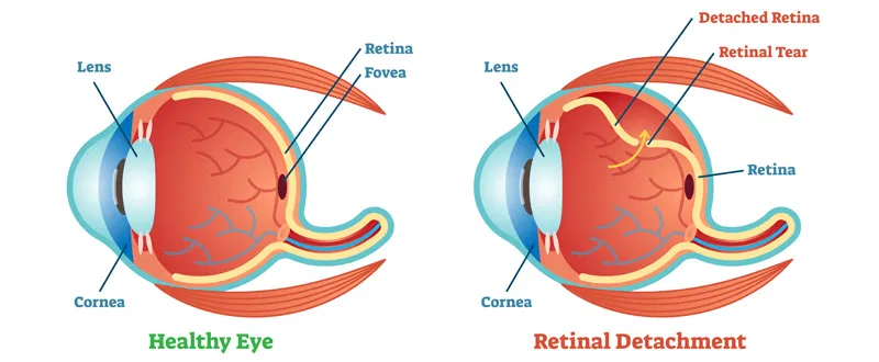
What Is Retinal Detachment?

Retinal detachment is a serious eye problem that may occur at any age although it usually occurs in middle-aged or older individuals. It is more likely to develop in people who are nearsighted (myopes) or in those whose relatives have had retinal detachments. A hard blow to the eye may cause the retina to detach as well. More rarely, this condition is a hereditary disease and may even occur in infants and children. If not treated early, retinal detachment may lead to total blindness in the affected eye.

The retina is a thin, transparent tissue of light-sensitive nerve fibres and cells, it covers the inside wall of the eye like wallpaper covers the walls of a room. The retinal functions like the film in a camera: light passes through the lens of the eye and is focused onto the retina. The light-sensitive retinal fibres are responsible for “taking the picture” and transmitting the image via the optic nerve to the brain.

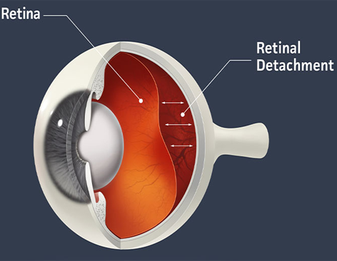
How Does Retinal Detachment Occur?

Most retinal detachments are caused by the presence of one or more small tears or holes in the retina. Normal aging can sometimes cause the retina to thin and degenerate, but more often shrinkage of the vitreous body-the clear gel-like substance which fills the centre of the eye, is responsible for causation of retinal tears.

Retinal tears are more common in myopic eyes and eyes with inflammation or injury. Once a retinal tear is present, watery fluid from the vitreous space may pass through the hole and flow between the retina and the back wall of the eye. This separates the retina from the back of the eye and causes it to ‘detach’. The part of the retina that is detached will not work properly and there will be a blur or a blind spot in vision.

Some retinal detachments are caused by other diseases in the eye such as tumours, severe inflammations, or complications of diabetes. These so-called secondary detachments do not have tears or holes in the retina and treatment of the disease which caused the retinal detachment can itself allow the retina to return to its normal position.

What Would Be the Symptoms?

Middle-aged and older persons may see floating black spots called floaters, and flashes of light in their vision. In most cases, these symptoms may not indicate serious problems, but in some eyes these symptoms may be associated with retinal tears. A comprehensive eye examination by an ophthalmologist to check the inside of the eye is necessary. Such an examination is desirable as soon as symptoms develop because fresh retinal tears before they lead to a more severe retinal detachment may be treatable without surgery.

Some retinal detachment patients may notice a wavy or watery quality in their overall vision or the appearance or a dark shadow in some part of their side vision. Further development of the retinal detachment will blur central vision and create significant sight loss in one eye unless the detachment is repaired. A few detachments may occur suddenly and the patient will experience a total loss of vision in one eye.

How to Diagnose Retinal Detachment?

A detached retina cannot be viewed from the outside of the eye therefore, if symptoms are noticed, an eye doctor having special training in retinal problems should be visited as soon as possible. He thoroughly examines the retina and the interior parts of the eye with an instrument called an indirect Ophthalmoscope. Other special diagnostic instruments including special contact lenses, slit lamp, and ultrasound may also be used.

How to Treat a Retinal Detachment?

There is no treatment with medicines for Retinal detachment. If the retina is tom and retinal detachment has not yet occurred a retinal detachment may be prevented by prompt prophylactic treatment. Once the retina becomes detached, it must be repaired surgically by an ophthalmologist. Successful re-attachment of the retina consists of sealing the retinal tear and preventing the retina from pulling away from the back of the eye again. There are several surgical procedures that may be used. The choice depends on the severity of the retinal detachment and the judgement of the ophthalmologist

Surgical Repair: The repair of retinal detachment may be approached by 3 different methods.

  1. Pneumatic Retinopexy: In a select group of cases, one may successfully attach the detached retina by injecting a gas bubble in the eye, treating the retinal hole with freezing (Cryo) or laser and positioning the eye to enable successful closure of the hole. This is the simplest of the treatment approaches with least intervention. The success of this procedure is about 70 per cent and in case of failure, the next procedure can be done with improved success (up to 90 per cent).
  2. Scleral Buckling: Most simple retinal detachments are handled by applying a silicone buckle on surface of the eye, thus indenting the walls inside. The retinal hole is treated with freezing (Cryo) and the fluid that has collected between retine and underlying layers is usually removed. The success of this surgery is usually very high, with almost 90 per cent of eyes having retinal reattachment with one procedure.
  3. Viteoretinal Procedures: For more complex retinal detachments, complicated surgery called vitreoretinal surgery is needed. In this, the diseased vitrous is removed along with abnormal scar tissue. The retina is attached by use of air, gas or silicone oil. The success of these surgeries varies with type of case. Sometimes, multiple surgeries may be indicated in case of recurrences. Where silicone oil is used, it is usually removed after variable period of time, once the retina is successfully reattached. The final success can be declared only if retina remains attached after removal of silicone oil. Obviously the more complex will be the surgical procedure needed and less will be the cure rate.

It is important to understand that surgical success and visual recovery need to go hand in hand. The visual recovery depends upon the basic strength in the retina, the duration of retinal detachment and most importantly the health of the central, most importantly the health of the central, most sensitive part of the retina called the macula. Reading fine print needs excellent vision, hence only a percentage of the eyes with complex retinal detachment can regain reading capabilities. More often, mobile vision is retrieved. Failed surgery usually leads to non recovery of vision and on occasions these eyes may shrink.

The surgery may be done under local or general anaesthesia, where gas/air silicone oil is injected the surgeon may advocate special posture. With gas in the eye, air travel is restricted Eye drops or ointment may have to be instilled for 6-8 weeks and glasses are prescribed at final examination. With simple buckling surgery, vision may start recovering in a few days time, but final vision is known after 6 weeks. With more complex vitreo-retinal surgeries, it takes longer time for visions to improve and stabilize.

In Summary

Patients with symptoms of retinal detachment require prompt attention by an ophthalmologist who will thoroughly examine the interior of the eye and advise about the need for treatment.

It is important for persons with significant myopia (near sightedness) or with family histories of retinal detachments to have periodic eye examinations by an ophthalmologist so that early detection of changes in the vitreous or retina may be diagnosed and potential retinal detachment prevented.

We, at Mansarovar are dedicated to the preservation and restoration of sight and are committed to providing the best possible care to your eyes, both now and in the future.

You must realise that every case is different and may not be the way it is described above. Hence, it may not answer all of your questions.

For comments and questions, click here.