Structure of the Eye
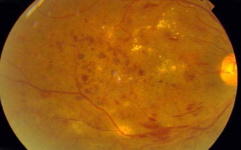
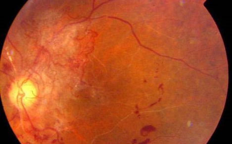
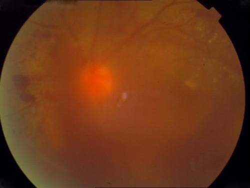
The inner most layer of the eye is composed of delicate tissue called the ‘retina’ which is comparable to the film inside a camera. Visual impulses are received by specialised cells in the retina and transmitted to the brain via the optic nerve. The central part of the retina which is the most sensitive and enables us to see clearly is called the ‘macula’.
What Are The Symptoms Of Diabetic Retinopathy?
How Is Diabetic Retinopathy Treated?
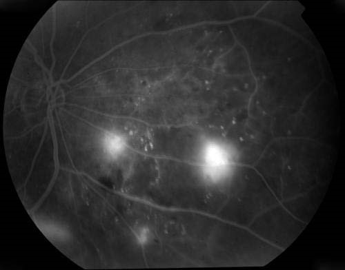
What Is Fundus Fluorescein Angiography – FFA Test?
This is a special test wherein a dye called ‘fluorescein’ is injected into the patient’s blood stream and once the dye reaches the retina pictures are taken of the retina using a special camera called ‘fundus camera’. This test has to be done in all patients undergoing laser treatment as it shows the doctor exactly what is the severity of the retinopathy. Also, when the test is repeated after laser treatment has been completed the effectiveness of the treatment can be evaluated and the film can be kept as a permanent record for future reference. All patients with significant retinopathy must periodically undergo this test.